Readings Newsletter
Become a Readings Member to make your shopping experience even easier.
Sign in or sign up for free!
You’re not far away from qualifying for FREE standard shipping within Australia
You’ve qualified for FREE standard shipping within Australia
The cart is loading…


This title is printed to order. This book may have been self-published. If so, we cannot guarantee the quality of the content. In the main most books will have gone through the editing process however some may not. We therefore suggest that you be aware of this before ordering this book. If in doubt check either the author or publisher’s details as we are unable to accept any returns unless they are faulty. Please contact us if you have any questions.
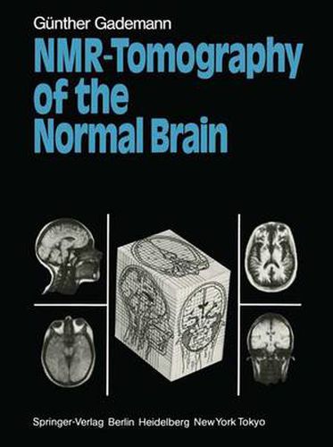
This book is intended as a short guide to the visualization of the anatomy of the normal brain by means of the NMR tomogram. The first section comprises a brief introduction to the physical and technical aspects of NMR. This is followed by the atlas section, which pursues a number of objectives. On the basis of two important NMR imaging techniques, the spin-echo technique and the inversion-recovery technique, those experi enced in CT are given the opportunity to familiarize themselves with the differences in tissue contrast that exist in NMR scans despite their appar ent similarity to conventional CT scans. The mode of action of the two NMR imaging techniques is explained in the technical introduction. An additional innovation is the possibility of producing sections that are not, as in CT scanning, limited by the body of the patient. The sagittal and frontal sections parallel to the plane of the face can show an unfamiliar, but particularly clear, image of the anatomy of the head and brain com pared with conventional horizontal sections. An anatomical description accompanying every section is provided by way of clarification. A particular advantage of NMR imaging, namely, the absence of ionizing radiation and, thus, an injurious effect on biological systems (Budinger 1981), makes it possible to provide a systematic visualization of a healthy human brain in a living person in three planes, arranged at right angles to one another.
$9.00 standard shipping within Australia
FREE standard shipping within Australia for orders over $100.00
Express & International shipping calculated at checkout
This title is printed to order. This book may have been self-published. If so, we cannot guarantee the quality of the content. In the main most books will have gone through the editing process however some may not. We therefore suggest that you be aware of this before ordering this book. If in doubt check either the author or publisher’s details as we are unable to accept any returns unless they are faulty. Please contact us if you have any questions.
This book is intended as a short guide to the visualization of the anatomy of the normal brain by means of the NMR tomogram. The first section comprises a brief introduction to the physical and technical aspects of NMR. This is followed by the atlas section, which pursues a number of objectives. On the basis of two important NMR imaging techniques, the spin-echo technique and the inversion-recovery technique, those experi enced in CT are given the opportunity to familiarize themselves with the differences in tissue contrast that exist in NMR scans despite their appar ent similarity to conventional CT scans. The mode of action of the two NMR imaging techniques is explained in the technical introduction. An additional innovation is the possibility of producing sections that are not, as in CT scanning, limited by the body of the patient. The sagittal and frontal sections parallel to the plane of the face can show an unfamiliar, but particularly clear, image of the anatomy of the head and brain com pared with conventional horizontal sections. An anatomical description accompanying every section is provided by way of clarification. A particular advantage of NMR imaging, namely, the absence of ionizing radiation and, thus, an injurious effect on biological systems (Budinger 1981), makes it possible to provide a systematic visualization of a healthy human brain in a living person in three planes, arranged at right angles to one another.
Search our extensive online catalogue.
