Readings Newsletter
Become a Readings Member to make your shopping experience even easier.
Sign in or sign up for free!
You’re not far away from qualifying for FREE standard shipping within Australia
You’ve qualified for FREE standard shipping within Australia
The cart is loading…


This title is printed to order. This book may have been self-published. If so, we cannot guarantee the quality of the content. In the main most books will have gone through the editing process however some may not. We therefore suggest that you be aware of this before ordering this book. If in doubt check either the author or publisher’s details as we are unable to accept any returns unless they are faulty. Please contact us if you have any questions.
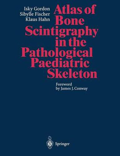
This very practical how-to guide comprehensively covers both the common and less common pathologies affecting the paediatric skeleton. It provides clear explanations of the materials and instrumentation, as well as teaching points, technical comments, discussions, and the avoidance of pitfalls. The images presented here have been produced using whole-body scanning, gamma-camera, high-resolution spot images, pinhole and SPECT, as well as three-phase bone scans - each procedure backed by indications for its use. These 350 illustrations thus allow the paediatrician, orthopaedic surgeon, radiologist and nuclear medicine physician a comparison with their own images as well as with the normal images presented in the authors’ companion volume, Atlas of Bone Scintigraphy in the Developing Paediatric Skeleton.
$9.00 standard shipping within Australia
FREE standard shipping within Australia for orders over $100.00
Express & International shipping calculated at checkout
This title is printed to order. This book may have been self-published. If so, we cannot guarantee the quality of the content. In the main most books will have gone through the editing process however some may not. We therefore suggest that you be aware of this before ordering this book. If in doubt check either the author or publisher’s details as we are unable to accept any returns unless they are faulty. Please contact us if you have any questions.
This very practical how-to guide comprehensively covers both the common and less common pathologies affecting the paediatric skeleton. It provides clear explanations of the materials and instrumentation, as well as teaching points, technical comments, discussions, and the avoidance of pitfalls. The images presented here have been produced using whole-body scanning, gamma-camera, high-resolution spot images, pinhole and SPECT, as well as three-phase bone scans - each procedure backed by indications for its use. These 350 illustrations thus allow the paediatrician, orthopaedic surgeon, radiologist and nuclear medicine physician a comparison with their own images as well as with the normal images presented in the authors’ companion volume, Atlas of Bone Scintigraphy in the Developing Paediatric Skeleton.