Readings Newsletter
Become a Readings Member to make your shopping experience even easier.
Sign in or sign up for free!
You’re not far away from qualifying for FREE standard shipping within Australia
You’ve qualified for FREE standard shipping within Australia
The cart is loading…


This title is printed to order. This book may have been self-published. If so, we cannot guarantee the quality of the content. In the main most books will have gone through the editing process however some may not. We therefore suggest that you be aware of this before ordering this book. If in doubt check either the author or publisher’s details as we are unable to accept any returns unless they are faulty. Please contact us if you have any questions.
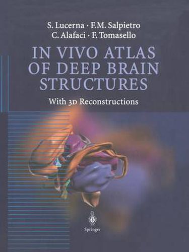
This ‘in vivo’ atlas contains more than 50 magnetic resonance (MR) images of the brain. Each structure is represented in the axial, coronal and sagittal plane, magnified in colour schemes and reconstructed in 3D images with a useful millimetric scale. The atlas offers the reader a practical and simple tool for surgical planning and for diagnostic and anatomical studies. The high level of anatomical definition of the in vivo MR images means that there is no loss in precision as a result of post-mortem changes. No doubt, this book is an excellent teaching instrument for all students of the neurosciences, regardless of the individual level of training and expertise.
$9.00 standard shipping within Australia
FREE standard shipping within Australia for orders over $100.00
Express & International shipping calculated at checkout
This title is printed to order. This book may have been self-published. If so, we cannot guarantee the quality of the content. In the main most books will have gone through the editing process however some may not. We therefore suggest that you be aware of this before ordering this book. If in doubt check either the author or publisher’s details as we are unable to accept any returns unless they are faulty. Please contact us if you have any questions.
This ‘in vivo’ atlas contains more than 50 magnetic resonance (MR) images of the brain. Each structure is represented in the axial, coronal and sagittal plane, magnified in colour schemes and reconstructed in 3D images with a useful millimetric scale. The atlas offers the reader a practical and simple tool for surgical planning and for diagnostic and anatomical studies. The high level of anatomical definition of the in vivo MR images means that there is no loss in precision as a result of post-mortem changes. No doubt, this book is an excellent teaching instrument for all students of the neurosciences, regardless of the individual level of training and expertise.