Readings Newsletter
Become a Readings Member to make your shopping experience even easier.
Sign in or sign up for free!
You’re not far away from qualifying for FREE standard shipping within Australia
You’ve qualified for FREE standard shipping within Australia
The cart is loading…


This title is printed to order. This book may have been self-published. If so, we cannot guarantee the quality of the content. In the main most books will have gone through the editing process however some may not. We therefore suggest that you be aware of this before ordering this book. If in doubt check either the author or publisher’s details as we are unable to accept any returns unless they are faulty. Please contact us if you have any questions.
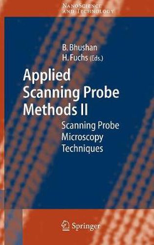
The Nobel Prize of 1986 on Sc- ningTunnelingMicroscopysignaled a new era in imaging. The sc- ning probes emerged as a new - strument for imaging with a p- cision suf?cient to delineate single atoms. At ?rst there were two - the Scanning Tunneling Microscope, or STM, and the Atomic Force Mic- scope, or AFM. The STM relies on electrons tunneling between tip and sample whereas the AFM depends on the force acting on the tip when it was placed near the sample. These were quickly followed by the M- netic Force Microscope, MFM, and the Electrostatic Force Microscope, EFM. The MFM will image a single magnetic bit with features as small as 10nm. With the EFM one can monitor the charge of a single electron. Prof. Paul Hansma at Santa Barbara opened the door even wider when he was able to image biological objects in aqueous environments. At this point the sluice gates were opened and a multitude of different instruments appeared. There are signi?cant differences between the Scanning Probe Microscopes or SPM, and others such as the Scanning Electron Microscope or SEM. The probe microscopes do not require preparation of the sample and they operate in ambient atmosphere, whereas, the SEM must operate in a vacuum environment and the sample must be cross-sectioned to expose the proper surface. However, the SEM can record 3D image and movies, features that are not available with the scanning probes.
$9.00 standard shipping within Australia
FREE standard shipping within Australia for orders over $100.00
Express & International shipping calculated at checkout
This title is printed to order. This book may have been self-published. If so, we cannot guarantee the quality of the content. In the main most books will have gone through the editing process however some may not. We therefore suggest that you be aware of this before ordering this book. If in doubt check either the author or publisher’s details as we are unable to accept any returns unless they are faulty. Please contact us if you have any questions.
The Nobel Prize of 1986 on Sc- ningTunnelingMicroscopysignaled a new era in imaging. The sc- ning probes emerged as a new - strument for imaging with a p- cision suf?cient to delineate single atoms. At ?rst there were two - the Scanning Tunneling Microscope, or STM, and the Atomic Force Mic- scope, or AFM. The STM relies on electrons tunneling between tip and sample whereas the AFM depends on the force acting on the tip when it was placed near the sample. These were quickly followed by the M- netic Force Microscope, MFM, and the Electrostatic Force Microscope, EFM. The MFM will image a single magnetic bit with features as small as 10nm. With the EFM one can monitor the charge of a single electron. Prof. Paul Hansma at Santa Barbara opened the door even wider when he was able to image biological objects in aqueous environments. At this point the sluice gates were opened and a multitude of different instruments appeared. There are signi?cant differences between the Scanning Probe Microscopes or SPM, and others such as the Scanning Electron Microscope or SEM. The probe microscopes do not require preparation of the sample and they operate in ambient atmosphere, whereas, the SEM must operate in a vacuum environment and the sample must be cross-sectioned to expose the proper surface. However, the SEM can record 3D image and movies, features that are not available with the scanning probes.