Readings Newsletter
Become a Readings Member to make your shopping experience even easier.
Sign in or sign up for free!
You’re not far away from qualifying for FREE standard shipping within Australia
You’ve qualified for FREE standard shipping within Australia
The cart is loading…


This title is printed to order. This book may have been self-published. If so, we cannot guarantee the quality of the content. In the main most books will have gone through the editing process however some may not. We therefore suggest that you be aware of this before ordering this book. If in doubt check either the author or publisher’s details as we are unable to accept any returns unless they are faulty. Please contact us if you have any questions.
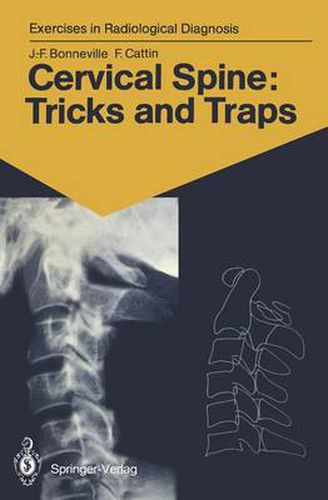
The cervical spine is always examined initially using standard radiographs, which often provide a sufficient basis for diagno- sis. Malformations, tumours, more frequently trauma, rheuma- tism, and even plain neck pain require a radiological exam- ination. The interpretation of radiological images is often difficult because of overlapping pieces of bone, the summation phe- nomena and the diversity of projections. In this book, two- or three-dimensional CTscans accompany the standard radiographs, serving as an excellent aid for com- prehension. It is almost as if the reader actually had the bones shown in the radiographs in his hands. From then on, everything becomes easy, superimpositions vanish, traps come to light, anatomy triumphs, and the images takes on life. Besanl$on, 1990 J .-F. BONNEVILLE, F. CATTIN v Contents Introduction 1 Iconography and Text with Corresponding Schemes 2 Subject Index …123 VII Normal cervical spine: 3-D imaging 1 1 2 Case 1 1 Our starting point is a normal lateral radiograph of the cervical spine. It will serve as a guide and point of reference in our task of comprehending the interrelationships between the structures of the cervical spine. This radiograph correctly includes the entire area from the base of the skull to the cervicothora- cic junction. The soft tissues are clearly visible anteriorly, as are the extremities of the spinous processes posteriorly. In this case, with the subject looking straight ahead, is a slight, regular, physiological lordosis ofthe cervical column.
$9.00 standard shipping within Australia
FREE standard shipping within Australia for orders over $100.00
Express & International shipping calculated at checkout
This title is printed to order. This book may have been self-published. If so, we cannot guarantee the quality of the content. In the main most books will have gone through the editing process however some may not. We therefore suggest that you be aware of this before ordering this book. If in doubt check either the author or publisher’s details as we are unable to accept any returns unless they are faulty. Please contact us if you have any questions.
The cervical spine is always examined initially using standard radiographs, which often provide a sufficient basis for diagno- sis. Malformations, tumours, more frequently trauma, rheuma- tism, and even plain neck pain require a radiological exam- ination. The interpretation of radiological images is often difficult because of overlapping pieces of bone, the summation phe- nomena and the diversity of projections. In this book, two- or three-dimensional CTscans accompany the standard radiographs, serving as an excellent aid for com- prehension. It is almost as if the reader actually had the bones shown in the radiographs in his hands. From then on, everything becomes easy, superimpositions vanish, traps come to light, anatomy triumphs, and the images takes on life. Besanl$on, 1990 J .-F. BONNEVILLE, F. CATTIN v Contents Introduction 1 Iconography and Text with Corresponding Schemes 2 Subject Index …123 VII Normal cervical spine: 3-D imaging 1 1 2 Case 1 1 Our starting point is a normal lateral radiograph of the cervical spine. It will serve as a guide and point of reference in our task of comprehending the interrelationships between the structures of the cervical spine. This radiograph correctly includes the entire area from the base of the skull to the cervicothora- cic junction. The soft tissues are clearly visible anteriorly, as are the extremities of the spinous processes posteriorly. In this case, with the subject looking straight ahead, is a slight, regular, physiological lordosis ofthe cervical column.
Search our extensive online catalogue.
