Readings Newsletter
Become a Readings Member to make your shopping experience even easier.
Sign in or sign up for free!
You’re not far away from qualifying for FREE standard shipping within Australia
You’ve qualified for FREE standard shipping within Australia
The cart is loading…


This title is printed to order. This book may have been self-published. If so, we cannot guarantee the quality of the content. In the main most books will have gone through the editing process however some may not. We therefore suggest that you be aware of this before ordering this book. If in doubt check either the author or publisher’s details as we are unable to accept any returns unless they are faulty. Please contact us if you have any questions.
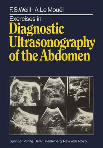
This book of diagnostic exercises cannot be used to good advantage without a good grasp of elementary sonoanatomy and the most common pathologic l images . We have tried to follow a pedagogical progression from the simple to the complicated for each group of clinical situations. We recommend that the sonograms at the beginning of each case study be thoroughly analysed before proceeding to the commentaries which explain the grounds for the final diagnosis. These explanatory remarks are accompanied by the same sonograms, but with arrows and letters added so as to pinpoint the details referred to as the diagnosis progresses. In reading the commentaries it will therefore be a good idea to cover over the figures in which the details are picked out for you, uncovering them one by one as required. 1 Which the reader may obtain from our previous books: Ed., 1982) Ultrasonography of Digestive Diseases (Mosby Publ., 2nd Renal Sonography (Springer Verlag, 1981) 1 Chapter 1 In Which the Reader is Invited to Clean His Glasses 1.1. Mrs. Beech, 75 years, has the complexion of a young girl, but she is losing weight and complains of epigastric pain. She has undergone a whole series of conventional radiological procedures; this may be good news for the film manufacturers, but it has not aided in the diagnosis. Finally, she is referred for an ultrasound examination. Look first at ultrasonic cuts 1.1a, b (transverse), then LId (sagittal).
$9.00 standard shipping within Australia
FREE standard shipping within Australia for orders over $100.00
Express & International shipping calculated at checkout
This title is printed to order. This book may have been self-published. If so, we cannot guarantee the quality of the content. In the main most books will have gone through the editing process however some may not. We therefore suggest that you be aware of this before ordering this book. If in doubt check either the author or publisher’s details as we are unable to accept any returns unless they are faulty. Please contact us if you have any questions.
This book of diagnostic exercises cannot be used to good advantage without a good grasp of elementary sonoanatomy and the most common pathologic l images . We have tried to follow a pedagogical progression from the simple to the complicated for each group of clinical situations. We recommend that the sonograms at the beginning of each case study be thoroughly analysed before proceeding to the commentaries which explain the grounds for the final diagnosis. These explanatory remarks are accompanied by the same sonograms, but with arrows and letters added so as to pinpoint the details referred to as the diagnosis progresses. In reading the commentaries it will therefore be a good idea to cover over the figures in which the details are picked out for you, uncovering them one by one as required. 1 Which the reader may obtain from our previous books: Ed., 1982) Ultrasonography of Digestive Diseases (Mosby Publ., 2nd Renal Sonography (Springer Verlag, 1981) 1 Chapter 1 In Which the Reader is Invited to Clean His Glasses 1.1. Mrs. Beech, 75 years, has the complexion of a young girl, but she is losing weight and complains of epigastric pain. She has undergone a whole series of conventional radiological procedures; this may be good news for the film manufacturers, but it has not aided in the diagnosis. Finally, she is referred for an ultrasound examination. Look first at ultrasonic cuts 1.1a, b (transverse), then LId (sagittal).
Search our extensive online catalogue.
