Readings Newsletter
Become a Readings Member to make your shopping experience even easier.
Sign in or sign up for free!
You’re not far away from qualifying for FREE standard shipping within Australia
You’ve qualified for FREE standard shipping within Australia
The cart is loading…


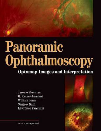
Panoramic Ophthalmoscopy: Optomap ® Images and Interpretation comprehensively covers the state-of-the-art technology and the high-resolution digital images taken with the Panoramic200 Scanning Laser Ophthalmoscope. The optomap ® Retinal Exam images provide ophthalmologists and optometrists with an extended view and photo-documentation of almost the entire retina.
Inside Panoramic Ophthalmoscopy, Jerome Sherman, Gulshan Karamchandani, William Jones, Sanjeev Nath, and Lawrence A. Yannuzzi document and expertly explain all there is to know about this remarkable new technology. Over 500 images highlight the text, many of which have never been seen before, and provide detailed visual references for numerous eye disorders.
This colorful atlas is the ideal resource for interpreting these images and diagnosing serious eye conditions that may have otherwise gone undetected.
Panoramic Ophthalmoscopy contains an introductory chapter that highlights and contrasts panoramic ophthalmoscopy and optomap ® images to all the traditional methods of fundus viewing. Inside you will find over 100 exemplary case presentations covering common and uncommon topics such as normal fundus, retinal tears, Coat’s disease, and diabetic retinopathy. Also included are cases of retinal and choroidal diseases and how they were diagnosed and managed using this technology. In the last chapter, the authors peer into the next frontier of imaging by introducing Optos fluorescein angiography and its myriad potential contributions to patient care, research, and clinical teaching.
Each case presentation includes:
History and chief compliant Clinical findings optomap ® images Differential diagnosis Disposition and follow-up
Cases are arranged into 11 chapters covering:
Optic Disc Macula Vascular Inflammatory Mass Lesions Retinal Degenerations Peripheral Lesions
With expert descriptions and hundreds of never before seen images, the all encompassing Panoramic Ophthalmoscopy: Optomap ® Images and Interpretation is the perfect resource for optometrists, ophthalmologists, ophthalmic technicians, residents, and students who would like to learn more about and would like to benefit from this revolutionary technology.
$9.00 standard shipping within Australia
FREE standard shipping within Australia for orders over $100.00
Express & International shipping calculated at checkout
Panoramic Ophthalmoscopy: Optomap ® Images and Interpretation comprehensively covers the state-of-the-art technology and the high-resolution digital images taken with the Panoramic200 Scanning Laser Ophthalmoscope. The optomap ® Retinal Exam images provide ophthalmologists and optometrists with an extended view and photo-documentation of almost the entire retina.
Inside Panoramic Ophthalmoscopy, Jerome Sherman, Gulshan Karamchandani, William Jones, Sanjeev Nath, and Lawrence A. Yannuzzi document and expertly explain all there is to know about this remarkable new technology. Over 500 images highlight the text, many of which have never been seen before, and provide detailed visual references for numerous eye disorders.
This colorful atlas is the ideal resource for interpreting these images and diagnosing serious eye conditions that may have otherwise gone undetected.
Panoramic Ophthalmoscopy contains an introductory chapter that highlights and contrasts panoramic ophthalmoscopy and optomap ® images to all the traditional methods of fundus viewing. Inside you will find over 100 exemplary case presentations covering common and uncommon topics such as normal fundus, retinal tears, Coat’s disease, and diabetic retinopathy. Also included are cases of retinal and choroidal diseases and how they were diagnosed and managed using this technology. In the last chapter, the authors peer into the next frontier of imaging by introducing Optos fluorescein angiography and its myriad potential contributions to patient care, research, and clinical teaching.
Each case presentation includes:
History and chief compliant Clinical findings optomap ® images Differential diagnosis Disposition and follow-up
Cases are arranged into 11 chapters covering:
Optic Disc Macula Vascular Inflammatory Mass Lesions Retinal Degenerations Peripheral Lesions
With expert descriptions and hundreds of never before seen images, the all encompassing Panoramic Ophthalmoscopy: Optomap ® Images and Interpretation is the perfect resource for optometrists, ophthalmologists, ophthalmic technicians, residents, and students who would like to learn more about and would like to benefit from this revolutionary technology.