Readings Newsletter
Become a Readings Member to make your shopping experience even easier.
Sign in or sign up for free!
You’re not far away from qualifying for FREE standard shipping within Australia
You’ve qualified for FREE standard shipping within Australia
The cart is loading…


Presents clinically applicable information about the use of cytology
Presents cases at the end of each chapter that help veterinarians appreciate the usefulness of cytology in ensuring a high quality diagnosis in their practice
Includes chapters written by experts from around the world
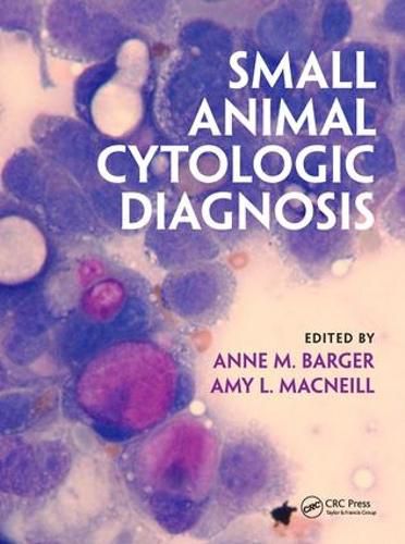
Contains more than 1300 superb illustrations. The colour schemes used throughout the book fit with the colours often seen with cytological staining, keeping the appearance of the book consistent, easy to look at and enjoyable to use. The book is divided up into chapters based on anatomical region, making it very easy to locate information required for assistance with a specific case. Tissue-specific chapters focus on diseases of a particular area, always in comparison to normal tissue. Unlike in other books, ocular and aural cytology hasn’t been grouped together as ‘organs of special senses’ and instead each have their own detailed chapters, providing a good breadth of information. Multiple cytological images are provided for the same sample, providing multiple views of what may be seen.
Summary tables give a quick reference that can be easily understood and used for real life scenarios.
The writing uses technical language where appropriate but without overcomplicating the information presented. Compared to other textbooks, this is very accessible and easy to understand.
The book is priced more affordably than the main competitor: Raskin & Meyer ‘Canine and Feline Cytology’ and it is more easily understandable, approachable, with more and better images.
$9.00 standard shipping within Australia
FREE standard shipping within Australia for orders over $100.00
Express & International shipping calculated at checkout
Presents clinically applicable information about the use of cytology
Presents cases at the end of each chapter that help veterinarians appreciate the usefulness of cytology in ensuring a high quality diagnosis in their practice
Includes chapters written by experts from around the world
Contains more than 1300 superb illustrations. The colour schemes used throughout the book fit with the colours often seen with cytological staining, keeping the appearance of the book consistent, easy to look at and enjoyable to use. The book is divided up into chapters based on anatomical region, making it very easy to locate information required for assistance with a specific case. Tissue-specific chapters focus on diseases of a particular area, always in comparison to normal tissue. Unlike in other books, ocular and aural cytology hasn’t been grouped together as ‘organs of special senses’ and instead each have their own detailed chapters, providing a good breadth of information. Multiple cytological images are provided for the same sample, providing multiple views of what may be seen.
Summary tables give a quick reference that can be easily understood and used for real life scenarios.
The writing uses technical language where appropriate but without overcomplicating the information presented. Compared to other textbooks, this is very accessible and easy to understand.
The book is priced more affordably than the main competitor: Raskin & Meyer ‘Canine and Feline Cytology’ and it is more easily understandable, approachable, with more and better images.