Readings Newsletter
Become a Readings Member to make your shopping experience even easier.
Sign in or sign up for free!
You’re not far away from qualifying for FREE standard shipping within Australia
You’ve qualified for FREE standard shipping within Australia
The cart is loading…


This title is printed to order. This book may have been self-published. If so, we cannot guarantee the quality of the content. In the main most books will have gone through the editing process however some may not. We therefore suggest that you be aware of this before ordering this book. If in doubt check either the author or publisher’s details as we are unable to accept any returns unless they are faulty. Please contact us if you have any questions.
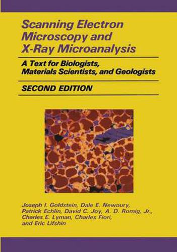
In the last decade, since the publication of the first edition of Scanning Electron Microscopy and X-ray Microanalysis, there has been a great expansion in the capabilities of the basic SEM and EPMA. High resolution imaging has been developed with the aid of an extensive range of field emission gun (FEG) microscopes. The magnification ranges of these instruments now overlap those of the transmission electron microscope. Low-voltage microscopy using the FEG now allows for the observation of noncoated samples. In addition, advances in the develop ment of x-ray wavelength and energy dispersive spectrometers allow for the measurement of low-energy x-rays, particularly from the light elements (B, C, N, 0). In the area of x-ray microanalysis, great advances have been made, particularly with the phi rho z [Ij)(pz)] technique for solid samples, and with other quantitation methods for thin films, particles, rough surfaces, and the light elements. In addition, x-ray imaging has advanced from the conventional technique of dot mapping to the method of quantitative compositional imaging. Beyond this, new software has allowed the development of much more meaningful displays for both imaging and quantitative analysis results and the capability for integrating the data to obtain specific information such as precipitate size, chemical analysis in designated areas or along specific directions, and local chemical inhomogeneities.
$9.00 standard shipping within Australia
FREE standard shipping within Australia for orders over $100.00
Express & International shipping calculated at checkout
This title is printed to order. This book may have been self-published. If so, we cannot guarantee the quality of the content. In the main most books will have gone through the editing process however some may not. We therefore suggest that you be aware of this before ordering this book. If in doubt check either the author or publisher’s details as we are unable to accept any returns unless they are faulty. Please contact us if you have any questions.
In the last decade, since the publication of the first edition of Scanning Electron Microscopy and X-ray Microanalysis, there has been a great expansion in the capabilities of the basic SEM and EPMA. High resolution imaging has been developed with the aid of an extensive range of field emission gun (FEG) microscopes. The magnification ranges of these instruments now overlap those of the transmission electron microscope. Low-voltage microscopy using the FEG now allows for the observation of noncoated samples. In addition, advances in the develop ment of x-ray wavelength and energy dispersive spectrometers allow for the measurement of low-energy x-rays, particularly from the light elements (B, C, N, 0). In the area of x-ray microanalysis, great advances have been made, particularly with the phi rho z [Ij)(pz)] technique for solid samples, and with other quantitation methods for thin films, particles, rough surfaces, and the light elements. In addition, x-ray imaging has advanced from the conventional technique of dot mapping to the method of quantitative compositional imaging. Beyond this, new software has allowed the development of much more meaningful displays for both imaging and quantitative analysis results and the capability for integrating the data to obtain specific information such as precipitate size, chemical analysis in designated areas or along specific directions, and local chemical inhomogeneities.