Readings Newsletter
Become a Readings Member to make your shopping experience even easier.
Sign in or sign up for free!
You’re not far away from qualifying for FREE standard shipping within Australia
You’ve qualified for FREE standard shipping within Australia
The cart is loading…


This title is printed to order. This book may have been self-published. If so, we cannot guarantee the quality of the content. In the main most books will have gone through the editing process however some may not. We therefore suggest that you be aware of this before ordering this book. If in doubt check either the author or publisher’s details as we are unable to accept any returns unless they are faulty. Please contact us if you have any questions.
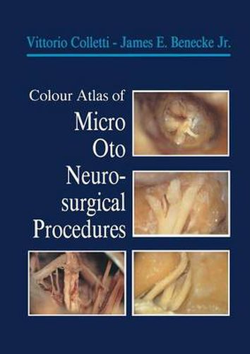
Modern microsurgical techniques have opened up a new horizon for the otoneurosurgeon. This volume is a very important contribu tion to the student who is learning these surgical approaches. Surgical otoneurology has now passed the infancy stage, but is still an adolescent. As more otologists and neurosurgeons become skilled in this type of surgery, new and better approaches will evolve. Certainly there needs to be much better management of the carotid artery as it passes through the temporal bone. Better techniques to preserve the IX, X, and XI nerves in the jugular bulb area should be developed, and more delicate procedures for management of lesions inside the cochlea and vestibular labyrinth should be developed. As our diagnostic techniques have improved, particularly through imaging, surgical techniques to match the improved diagnostic techniques will emerge. For future otoneurologists who are pre pared, many problems involving the temporal bone that are now considered untreatable will be successfully managed for very grateful patients. The purpose of this text is to familiarize the otoneurosur geon with the anatomy of the temporal bone, skull base, infratem poral fossa, and cerebellopontine angle. This anatomy will be taught by demonstrating surgical procedures. This atlas which is an example of cooperation between the schools of Los Angeles and Verona will permit the reader to rehearse otoneurosurgical procedures in the laboratory, and, when the techniques have been mastered, apply the various approaches in the treatment of inner ear and skull base lesions. William F. House MD.
$9.00 standard shipping within Australia
FREE standard shipping within Australia for orders over $100.00
Express & International shipping calculated at checkout
This title is printed to order. This book may have been self-published. If so, we cannot guarantee the quality of the content. In the main most books will have gone through the editing process however some may not. We therefore suggest that you be aware of this before ordering this book. If in doubt check either the author or publisher’s details as we are unable to accept any returns unless they are faulty. Please contact us if you have any questions.
Modern microsurgical techniques have opened up a new horizon for the otoneurosurgeon. This volume is a very important contribu tion to the student who is learning these surgical approaches. Surgical otoneurology has now passed the infancy stage, but is still an adolescent. As more otologists and neurosurgeons become skilled in this type of surgery, new and better approaches will evolve. Certainly there needs to be much better management of the carotid artery as it passes through the temporal bone. Better techniques to preserve the IX, X, and XI nerves in the jugular bulb area should be developed, and more delicate procedures for management of lesions inside the cochlea and vestibular labyrinth should be developed. As our diagnostic techniques have improved, particularly through imaging, surgical techniques to match the improved diagnostic techniques will emerge. For future otoneurologists who are pre pared, many problems involving the temporal bone that are now considered untreatable will be successfully managed for very grateful patients. The purpose of this text is to familiarize the otoneurosur geon with the anatomy of the temporal bone, skull base, infratem poral fossa, and cerebellopontine angle. This anatomy will be taught by demonstrating surgical procedures. This atlas which is an example of cooperation between the schools of Los Angeles and Verona will permit the reader to rehearse otoneurosurgical procedures in the laboratory, and, when the techniques have been mastered, apply the various approaches in the treatment of inner ear and skull base lesions. William F. House MD.