Readings Newsletter
Become a Readings Member to make your shopping experience even easier.
Sign in or sign up for free!
You’re not far away from qualifying for FREE standard shipping within Australia
You’ve qualified for FREE standard shipping within Australia
The cart is loading…


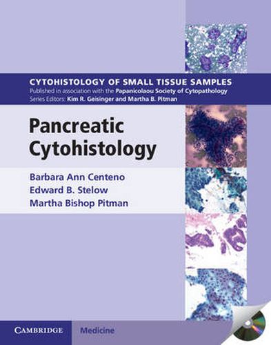
Each volume in this richly illustrated series, sponsored by the Papanicolaou Society of Cytopathology, provides an organ-based approach to the cytologic and histologic diagnosis of small tissue samples including fine-needle aspiration biopsy, cell block samples and core, pinch and forceps biopsies. This volume provides a practical approach to preparing and assessing pancreatic aspiration, core biopsy and brushing samples. Benign, pre-malignant and malignant entities are presented in a well-organized and standardized format supported with high-resolution color photomicrographs, tables, tabulated specific morphologic criteria and appropriate ancillary testing algorithms. Example vignettes allow the reader to assimilate the diagnostic principles in a case-based format. This unique series strengthens the bridge between surgical pathology and cytopathology, providing the pathologist with the ability to diagnose small tissue samples with confidence. The CD-ROM packaged with the printed book contains all the images in a downloadable format, making this a valuable resource for practicing and trainee pathologists.
$9.00 standard shipping within Australia
FREE standard shipping within Australia for orders over $100.00
Express & International shipping calculated at checkout
Each volume in this richly illustrated series, sponsored by the Papanicolaou Society of Cytopathology, provides an organ-based approach to the cytologic and histologic diagnosis of small tissue samples including fine-needle aspiration biopsy, cell block samples and core, pinch and forceps biopsies. This volume provides a practical approach to preparing and assessing pancreatic aspiration, core biopsy and brushing samples. Benign, pre-malignant and malignant entities are presented in a well-organized and standardized format supported with high-resolution color photomicrographs, tables, tabulated specific morphologic criteria and appropriate ancillary testing algorithms. Example vignettes allow the reader to assimilate the diagnostic principles in a case-based format. This unique series strengthens the bridge between surgical pathology and cytopathology, providing the pathologist with the ability to diagnose small tissue samples with confidence. The CD-ROM packaged with the printed book contains all the images in a downloadable format, making this a valuable resource for practicing and trainee pathologists.