Readings Newsletter
Become a Readings Member to make your shopping experience even easier.
Sign in or sign up for free!
You’re not far away from qualifying for FREE standard shipping within Australia
You’ve qualified for FREE standard shipping within Australia
The cart is loading…


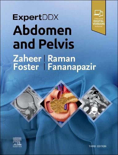
Highly practical and user-friendly, ExpertDDx: Abdomen and Pelvis, third edition, helps you reach accurate, clinically useful differential diagnoses in your everyday practice. It presents the most useful differential diagnoses for each region of the abdomen and pelvis, grouped according to anatomic location, generic imaging findings, modality-specific findings, or clinical-based indications. Each differential diagnosis includes several high-quality, succinctly annotated images; a list of diagnostic possibilities sorted as common, less common, and rare but important; and brief, bulleted text offering helpful diagnostic clues. It’s an excellent resource for subspecialty abdominal imagers as well as general radiologists and trainees, providing invaluable assistance in reaching logical, on-target differential diagnoses based on key imaging findings and clinical details.
Covers 175 of the most common diagnostic challenges in abdominal and pelvic imaging, enhanced by more than 2,100 radiologic images, full-color illustrations, clinical and histologic photographs, and gross pathology images
Provides a quick review of the salient features of each entity, differentiating features from other similar-appearing abnormalities
Includes new chapters on hematuria, flank pain, acute scrotal pain, and seminal vesicle
Adds greater focus to advancing prostate imaging methods with expanded content on lesions in the peripheral zone and lesions in the transition zone, as well as new coverage of transplant imaging
Contains updates to numerous classifications, including LI-RADS for liver, O-RADS for ovarian masses, and the Tanaka classification for pancreatic cysts
Features new MR examples and MR-specific diagnoses throughout, plus new differentials for contrast-enhanced ultrasound findings related to liver and kidney lesions
Includes the enhanced eBook version, which allows you to search all text, figures, and references on a variety of devices
$9.00 standard shipping within Australia
FREE standard shipping within Australia for orders over $100.00
Express & International shipping calculated at checkout
Highly practical and user-friendly, ExpertDDx: Abdomen and Pelvis, third edition, helps you reach accurate, clinically useful differential diagnoses in your everyday practice. It presents the most useful differential diagnoses for each region of the abdomen and pelvis, grouped according to anatomic location, generic imaging findings, modality-specific findings, or clinical-based indications. Each differential diagnosis includes several high-quality, succinctly annotated images; a list of diagnostic possibilities sorted as common, less common, and rare but important; and brief, bulleted text offering helpful diagnostic clues. It’s an excellent resource for subspecialty abdominal imagers as well as general radiologists and trainees, providing invaluable assistance in reaching logical, on-target differential diagnoses based on key imaging findings and clinical details.
Covers 175 of the most common diagnostic challenges in abdominal and pelvic imaging, enhanced by more than 2,100 radiologic images, full-color illustrations, clinical and histologic photographs, and gross pathology images
Provides a quick review of the salient features of each entity, differentiating features from other similar-appearing abnormalities
Includes new chapters on hematuria, flank pain, acute scrotal pain, and seminal vesicle
Adds greater focus to advancing prostate imaging methods with expanded content on lesions in the peripheral zone and lesions in the transition zone, as well as new coverage of transplant imaging
Contains updates to numerous classifications, including LI-RADS for liver, O-RADS for ovarian masses, and the Tanaka classification for pancreatic cysts
Features new MR examples and MR-specific diagnoses throughout, plus new differentials for contrast-enhanced ultrasound findings related to liver and kidney lesions
Includes the enhanced eBook version, which allows you to search all text, figures, and references on a variety of devices