Readings Newsletter
Become a Readings Member to make your shopping experience even easier.
Sign in or sign up for free!
You’re not far away from qualifying for FREE standard shipping within Australia
You’ve qualified for FREE standard shipping within Australia
The cart is loading…


For two-semester A&P lab courses. Stunning Visuals and Accessible Tutorials Engage Students in the A&P Lab
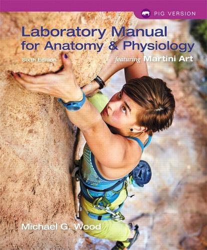
The Laboratory Manual for Anatomy & Physiology featuring Martini Art, is a valuable resource for engaging students in the lab, introducing them to applications, and preparing them for their future careers. This edition teaches effective drawing techniques to promote critical thinking and ensure lasting comprehension. This comprehensive lab manual features more than 100 new photos that walk students through core lab processes, lab equipment, and animal organ dissections, as well as art that is adapted from Martini’s Fundamentals of Anatomy & Physiology. It is available in three formats: Main, Cat, and Pig Versions. The Cat and Pig manuals are identical to the Main Version, with nine additional cat or pig dissection exercises.
Features
Draw It! Tutorials show the students how to quickly and easily draw select structures and processes to better understand them. Pictorial instruction photos walk students through the steps of core lab processes and standard procedures in the field, such as how to examine microscopic structures. Model Photos are clear, precise anatomical model photos provided by the author. Stepwise Dissection Photos visually guide students through examinations of animal organs Lab Equipment and Clinical Photos effectively train students how to work in labs and professional clinical settings. Photo topics include:
Students using key lab equipment Clinical instruments and medical devices Clinicians performing procedures
Drawing boxes are modifiable to enlarge and match the instructor’s preferred look for the final assignment.
Martini Art brings together a comprehensive program featuring histology micrographs by Michael Wood, and high-quality cat and pig dissection photos by the expert dissector and photographer team of Shawn Miller and Mark Nielsen at the University of Utah.
$9.00 standard shipping within Australia
FREE standard shipping within Australia for orders over $100.00
Express & International shipping calculated at checkout
For two-semester A&P lab courses. Stunning Visuals and Accessible Tutorials Engage Students in the A&P Lab
The Laboratory Manual for Anatomy & Physiology featuring Martini Art, is a valuable resource for engaging students in the lab, introducing them to applications, and preparing them for their future careers. This edition teaches effective drawing techniques to promote critical thinking and ensure lasting comprehension. This comprehensive lab manual features more than 100 new photos that walk students through core lab processes, lab equipment, and animal organ dissections, as well as art that is adapted from Martini’s Fundamentals of Anatomy & Physiology. It is available in three formats: Main, Cat, and Pig Versions. The Cat and Pig manuals are identical to the Main Version, with nine additional cat or pig dissection exercises.
Features
Draw It! Tutorials show the students how to quickly and easily draw select structures and processes to better understand them. Pictorial instruction photos walk students through the steps of core lab processes and standard procedures in the field, such as how to examine microscopic structures. Model Photos are clear, precise anatomical model photos provided by the author. Stepwise Dissection Photos visually guide students through examinations of animal organs Lab Equipment and Clinical Photos effectively train students how to work in labs and professional clinical settings. Photo topics include:
Students using key lab equipment Clinical instruments and medical devices Clinicians performing procedures
Drawing boxes are modifiable to enlarge and match the instructor’s preferred look for the final assignment.
Martini Art brings together a comprehensive program featuring histology micrographs by Michael Wood, and high-quality cat and pig dissection photos by the expert dissector and photographer team of Shawn Miller and Mark Nielsen at the University of Utah.