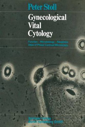
Gynecological Vital Cytology: Function - Microbiology - Neoplasia Atlas of Phase-Contrast Microscopy
Peter Stoll,Gisela Dallenbach-Hellweg,Peter Stoll,Gisela Dallenbach-Hellweg

Gynecological Vital Cytology: Function - Microbiology - Neoplasia Atlas of Phase-Contrast Microscopy
Peter Stoll,Gisela Dallenbach-Hellweg,Peter Stoll,Gisela Dallenbach-Hellweg
This title is printed to order. This book may have been self-published. If so, we cannot guarantee the quality of the content. In the main most books will have gone through the editing process however some may not. We therefore suggest that you be aware of this before ordering this book. If in doubt check either the author or publisher’s details as we are unable to accept any returns unless they are faulty. Please contact us if you have any questions.
In gynecological practice, techniques of examination are being supple- mented more and more by cytodiagnosis. Thus it has become necessary to acquaint the gynecologist of the possibilities, use and Iimits of cyto- diagnosis. Such is the purpose of the book Gynakologische Cytologie (Stall, Jaeger, Dallenbach, Springer-Verlag 1968). In general, the practicing gynecologist will merely make the vaginal, ecto-and endocervical smears and leave the diagnosis of them to a cyto- logical laboratory. Only in rare cases will a trained and experienced specialist set up his own cytologicallaboratory for outpatients, although such undertaking would be very desirable for propagating the cyto- logical method. The cytological analysis of unstained fresh smears during the gyne- cological examination allows an immediate study to be made of micro- f!ora and cellular atypia. F or such cytological studies microscopes are employed in which a high-cantrast image of the specimen is obtained by optical means (phase-contrast and interference-contrast microscopy), thereby eliminating the need for fixation and staining. In the nineteen-thirties the Dutch physicist Zernike investigated the formation of high-cantrast images of transparent objects by modifying the path of light. In 1941, his ideas were put into practice by the firm of Carl Zeiss, Jena. Zernike received the Nobel Prize for physics in 1953. The method he had developed proved of great value in biology and in medicine, above all for the examination of living objects. It was intro- duced into gynecology by Runge, Voege, Haselmann and Zinser in 1949.
This item is not currently in-stock. It can be ordered online and is expected to ship in 7-14 days
Our stock data is updated periodically, and availability may change throughout the day for in-demand items. Please call the relevant shop for the most current stock information. Prices are subject to change without notice.
Sign in or become a Readings Member to add this title to a wishlist.


